• Blog
  • About
  • Contact
  • Blog
  • About
  • Contact
Latrice Brentnall

Fallen Arches Causes, Signs Or Symptoms And Treatment

7/2/2017

 
Overview

Adult Acquired Flat Feet

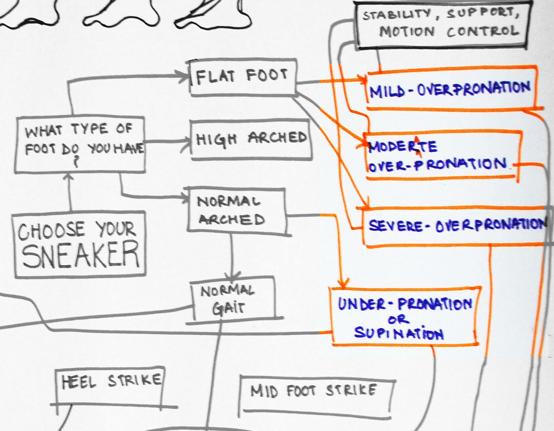
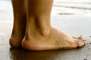
?Pes Planus? is the medical term for flat feet. It comes from Latin: Pes = foot and Planus = plain, level ground. Very few people suffer from this condition, as a true flat foot is very rare. Less than 5% of the population has flat feet. The majority of the Australian population, however, has fallen arches (an estimated 60-70% of the population) known in the medical profession as ?excess pronation? or over-pronation. Over-pronation means the foot and ankle tend to roll inwards and the arch collapses with weight-bearing. This is a quite a destructive position for the foot to function in and may cause a wide variety of foot, leg and lower back conditions.

Causes

There are several reasons why arches fall. It can be genetic, it can be a result of unsupported shoes, a product of our lives and the pressure we put on the feet. Some people have active lifestyles so the wear and tear effect on the muscles there that runs down the back of the calf and wraps around the arch can slacken as we get older. It can be a number of things.

Symptoms

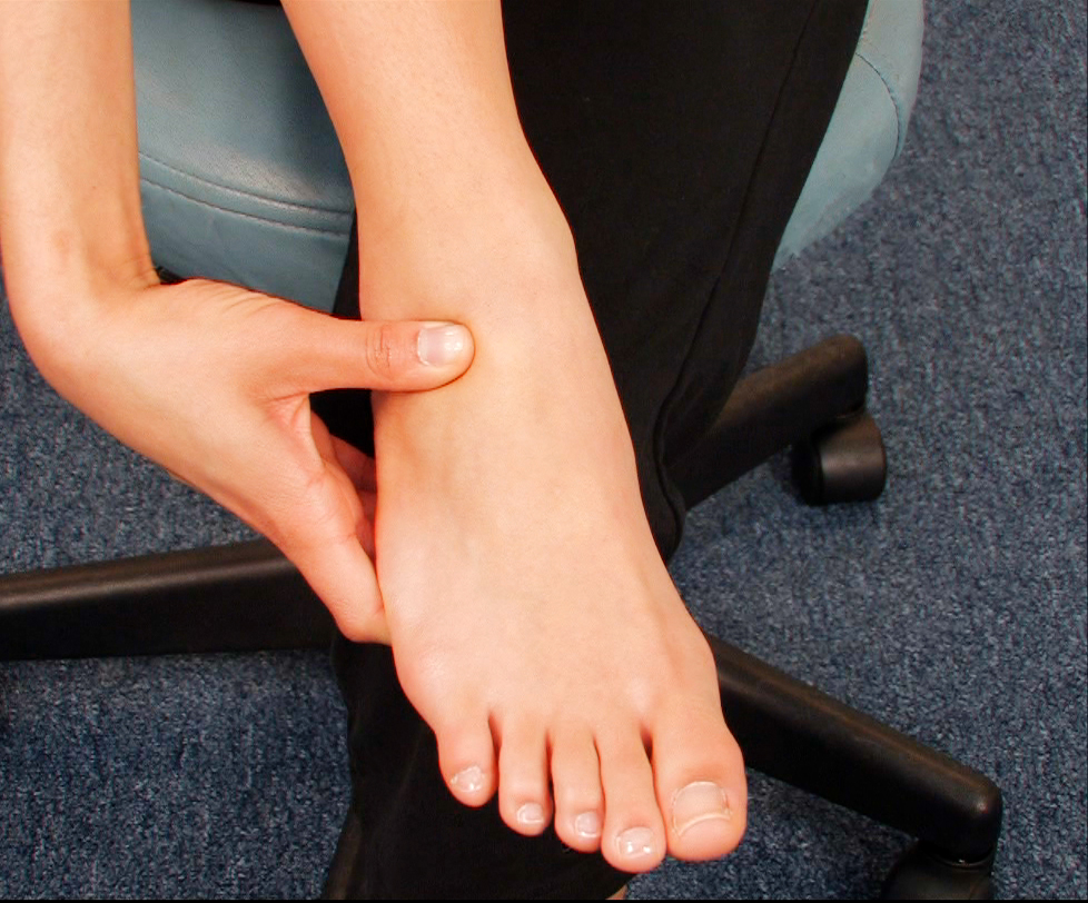
Feet tire easily and become painful and achy, especially around the arch, ankle and heel. Swelling on the inside bottom of your feet. Back and leg pain. Difficulty standing on toes.

Diagnosis

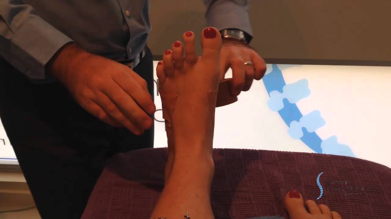
Your doctor will ask about your symptoms and medical history. A physical and foot exam will be done. Flat feet can be diagnosed by appearance. To determine if the foot is rigid, you may be asked to do some simple tasks.
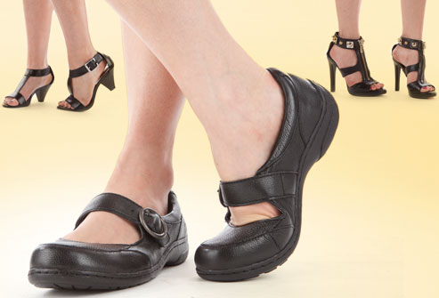
fallen arches shoes

Non Surgical Treatment

In rare cases, surgery may be needed if a child has flat feet caused by a problem they're born with (a congenital abnormality). The foot may need to be straightened or the bones may need to be separated if they're fused together. Painkillers and insoles are the first treatment options for flat feet that are caused by a joint problem, such as arthritis or a torn tendon. However, surgery may be recommended if the injury or condition is severely affecting your feet. Where flat feet are caused by a condition that affects the nervous system, special shoes, insoles, or supportive foot or leg braces may be needed. Again, in severe cases, an operation may be needed to straighten the feet.

Surgical Treatment

Flat Foot

Surgery for flat feet is separated into three kinds: soft tissue procedures, bone cuts, and bone fusions. Depending on the severity of the flat foot, a person?s age, and whether or not the foot is stiff determines just how the foot can be fixed. In most cases a combination of procedures are performed. With flexible flat feet, surgery is geared at maintaining the motion of the foot and recreating the arch. Commonly this may involve tendon repairs along the inside of the foot to reinforce the main tendon that lifts the arch. When the bone collapse is significant, bone procedures are included to physically rebuild the arch, and realign the heel. The presence of bunions with flat feet is often contributing to the collapse and in most situations requires correction. With rigid flat feet, surgery is focused on restoring the shape of the foot through procedures that eliminate motion. In this case, motion does not exist pre-operatively, so realigning the foot is of utmost importance. The exception, are rigid flat feet due to tarsal coalition (fused segment of bone) in the back of the foot where freeing the blockage can restore function.

After Care

Patients may go home the day of surgery or they may require an overnight hospital stay. The leg will be placed in a splint or cast and should be kept elevated for the first two weeks. At that point, sutures are removed. A new cast or a removable boot is then placed. It is important that patients do not put any weight on the corrected foot for six to eight weeks following the operation. Patients may begin bearing weight at eight weeks and usually progress to full weightbearing by 10 to 12 weeks. For some patients, weightbearing requires additional time. After 12 weeks, patients commonly can transition to wearing a shoe. Inserts and ankle braces are often used. Physical therapy may be recommended. There are complications that relate to surgery in general. These include the risks associated with anesthesia, infection, damage to nerves and blood vessels, and bleeding or blood clots. Complications following flatfoot surgery may include wound breakdown or nonunion (incomplete healing of the bones). These complications often can be prevented with proper wound care and rehabilitation. Occasionally, patients may notice some discomfort due to prominent hardware. Removal of hardware can be done at a later time if this is an issue. The overall complication rates for flatfoot surgery are low.

Functional Leg Length Discrepancy Running

6/30/2017

 
Overview

While many people have slight differences in the length of one leg compared to the other, a significant difference between the two legs can cause problems. Leg length discrepancies can disturb the way a person walks, how his or her spine lines up and strain hip joints.Leg Length Discrepancy

Causes

Some causes of leg length discrepancy (other than anatomical). Dysfunction of the hip joint itself leading to compensatory alterations by the joint and muscles that impact on the joint. Muscle mass itself, i.e., the vastus lateralis muscle, pushes the iliotibial band laterally, causing femoral compensations to maintain a line of progression during the gait cycle. This is often misdiagnosed as I-T band syndrome and subsequently treated incorrectly. The internal rotators of the lower limb are being chronically short or in a state of contracture. According to Cunningham's Manual of Practical Anatomy these are muscles whose insertion is lateral to the long axis of the femur. The external rotators of the hip joint are evidenced in the hip rotation test. The iliosacral joint displays joint fixations on the superior or inferior transverse, or the sagittal axes. This may result from many causes including joint, muscle, osseous or compensatory considerations. Short hamstring muscles, i.e., the long head of the biceps femoris muscle. In the closed kinetic chain an inability of the fibula to drop inferior will result in sacrotuberous ligament loading failure. The sacroiliac joint dysfunctions along its right or left oblique axis. Failure or incorrect loading of the Back Force Transmission System (the longitudinal-muscle-tendon-fascia sling and the oblique dorsal muscle-fascia-tendon sling). See the proceedings of the first and second Interdisciplinary World Congress on Low Back Pain. Sacral dysfunction (nutation or counternutation) on the respiratory axis. When we consider the above mentioned, and other causes, it should be obvious that unless we look at all of the causes of leg length discrepancy/asymmetry then we will most assuredly reach a diagnosis based on historical dogma or ritual rather than applying the rules of current differential diagnosis.

Symptoms

As patients develop LLD, they will naturally and even unknowingly attempt to compensate for the difference between their two legs by either bending the longer leg excessively or standing on the toes of the short leg. When walking, they are forced to step down on one side and thrust upwards on the other side, which leads to a gait pattern with an abnormal up and down motion. For many patients, especially adolescents, the appearance of their gait may be more personally troublesome than any symptoms that arise or any true functional deficiency. Over time, standing on one's toes can create a contracture at the ankle, in which the calf muscle becomes abnormally contracted, a condition that can help an LLD patient with walking, but may later require surgical repair. If substantial enough, LLD left untreated can contribute to other serious orthopaedic problems, such as degenerative arthritis, scoliosis, or lower back pain. However, with proper treatment, children with leg length discrepancy generally do quite well, without lingering functional or cosmetic deficiencies.

Diagnosis

Infants, children or adolescents suspected of having a limb-length condition should receive an evaluation at the first sign of difficulty in using their arms or legs. In many cases, signs are subtle and only noticeable in certain situations, such as when buying clothing or playing sports. Proper initial assessments by qualified pediatric orthopedic providers can reduce the likelihood of long-term complications and increase the likelihood that less invasive management will be effective. In most cases, very mild limb length discrepancies require no formal treatment at all.

Non Surgical Treatment

The non-surgical intervention is mainly usedfor the functional and environmental types of leg length discrepancies. It is also applied to the mild category of limb length inequality. Non-surgical intervention consists of stretching the muscles of the lower extremity. This is individually different, whereby the M. Tensor Fascia latae, the adductors, the hamstring muscles, M. piriformis and M. Iliopsoas are stretched. In this non-surgical intervention belongs also the use of shoe lifts. These shoe lifts consists of either a shoe insert (up to 10-20mm of correction), or building up the sole of the shoe on the shorter leg (up to 30-60mm of correction). This lift therapy should be implemented gradually in small increments. Several studies have examined the treatment of low back pain patients with LLD with shoe lifts. Gofton obtained good results: the patients experienced major or complete pain relief that lasted upon follow-up ranging from 3 to 11 years. Helliwell also observed patients whereby 44% experienced complete pain relief, and 45% had moderate or substantial pain relief. Friberg found that 157 (of 211) patients with LBP, treated with shoe lifts, were symprom-free after a mean follow-up of 18 months.

LLL Shoe Insoles

leg length discrepancy treatment adults

Surgical Treatment

Surgical treatments vary in complexity. Sometimes the goal of surgery is to stop the growth of the longer limb. Other times, surgeons work to lengthen the shorter limb. Orthopedic surgeons may treat children who have limb-length conditions with one or a combination of these surgical techniques. Bone resection. An operation to remove a section of bone, evening out the limbs in teens or adults who are no longer growing. Epiphyseal stapling. An operation to slow the rate of growth of the longer limb by inserting staples into the growth plate, then removing them when the desired result is achieved. Epiphysiodesis. An operation to slow the rate of growth of the longer limb by creating a permanent bony ridge near the growth plate. Limb lengthening. A procedure (also called distraction osteogenesis or the Ilizarov procedure) that involves attaching an internal or external fixator to a limb and gradually pulling apart bone segments to grow new bone between them. There are several ways your doctor can predict the final LLD, and thus the timing of the surgery. The easiest way is the so-called Australian method, popularised by Dr. Malcolm Menelaus, an Australian orthopedic surgeon. According to this method, growth in girls is estimated to stop at age 14, and in boys at age 16 years. The femur grows at the rate of 10 mm. a year, and the upper tibia at the rate of 6 mm. a year. Using simple arithmetic, one can get a fairly good prediction of future growth. This of course, is an average, and the patient may be an average. To cut down the risk of this, the doctor usually measures leg length using special X-ray technique (called a Scanogram) on three occasions over at least one year duration to estimate growth per year. He may also do an X-ray of the left hand to estimate the bone age (which in some cases may differ from chronological age) by comparing it with an atlas of bone age. In most cases, however, the bone age and chronological age are quite close. Another method of predicting final LLD is by using Anderson and Green?s remaining growth charts. This is a very cumbersome method, but was till the 1970?s, the only method of predicting remaining growth. More recently, however, a much more convenient method of predicting LLD was discovered by Dr. Colin Moseley from Montreal. His technique of using straight line graphs to plot growth of leg lengths is now the most widely used method of predicting leg length discrepancy. Whatever method your doctor uses, over a period of one or two years, once he has a good idea of the final LLD, he can then formulate a plan to equalize leg lengths. Epiphyseodesis is usually done in the last 2 to 3 years of growth, giving a maximum correction of about 5 cm. Leg lengthening can be done at any age, and can give corrections of 5 to10 cm., or more.

What Are The Major Causes Of Heel Pain

6/28/2017

 
Overview

Foot Pain

Heel pain is generally the result of faulty biomechanics (walking gait abnormalities) that place too much stress on the heel bone and the soft tissues that attach to it. The stress may also result from injury, or a bruise incurred while walking, running, or jumping on hard surfaces; wearing poorly constructed footwear; or being overweight. The heel bone is the largest of the 26 bones in the human foot, which also has 33 joints and a network of more than 100 tendons, muscles, and ligaments. Like all bones, it is subject to outside influences that can affect its integrity and its ability to keep us on our feet. Heel pain, sometimes disabling, can occur in the front, back, or bottom of the heel.

Causes

Here are a few in-depth explanations of heel pain causes and potential remedies for those that are afflicted Plantar Fascitis, One of the most well-known causes of heel pain, plantar fascitis occurs when the thick tissue along the arch of the foot becomes tight and inflamed. The foot itself can feel cramped when this tissue is inflamed, causing the afflicted person to feel as if they have a muscle cramp that flexing the foot doesn?t seem to alleviate. Plantar fascitis is most common among men over the age of 40. Warning Signs, The first steps in the morning are extremely painful. Pain flares up after activities like stair or steep hill climbing. Pain continues to occur regardless of the type of shoes or lack of shoes worn.

Symptoms

The symptoms of plantar fasciitis include pain along the inside edge of the heel near the arch of the foot. The pain is worse when weight is placed on the foot especially after a long period of rest or inactivity. This is usually most pronounced in the morning when the foot is first placed on the floor. This symptom called first-step pain is typical of plantar fasciitis. Prolonged standing can also increase the painful symptoms. It may feel better after activity but most patients report increased pain by the end of the day. Pressing on this part of the heel causes tenderness. Pulling the toes back toward the face can be very painful.

Diagnosis

The diagnosis of heel pain and heel spurs is made by a through history of the course of the condition and by physical exam. Weight bearing x-rays are useful in determining if a heel spur is present and to rule out rare causes of heel pain such as a stress fracture of the heel bone, the presence of bone tumors or evidence of soft tissue damage caused by certain connective tissue disorders.

Non Surgical Treatment

Treatment for plantar fasciitis - the vast majority of patients recover with conservative treatments (designed to avoid radical medical therapeutic measures or operative procedures) within months. Heel with ice-pack. Home care such as rest, ice-pack use, proper-fitting footwear and foot supports are often enough to ease heel pain. Non-steroidal anti-inflammatory drugs (NSAIDs) - medications with analgesic (pain reducing), antipyretic (fever reducing) effects. In higher doses they also have anti-inflammatory effects, they reduce inflammation (swelling). Non-steroidal distinguishes NSAIDs from other drugs which contain steroids, which are also anti-inflammatory. NSAIDs are non-narcotic (they do not induce stupor). For patients with plantar fasciitis they may help with pain and inflammation. Corticosteroids, a corticosteroid solution is applied over the affected area on the skin; an electric current is used to help absorption. Alternatively, the doctor may decide to inject the medication. However, multiple injections may result in a weakened plantar fascia, significantly increasing the risk of rupture and shrinkage of the fat pad covering the heel bone. Some doctors may use ultrasound to help them make sure they have injected in the right place Corticosteroids are usually recommended when NSAIDs have not helped. Physical therapy, a qualified/specialized physical therapist (UK: physiotherapist) can teach the patient exercises which stretch the plantar fascia and Achilles tendon, as well as strengthening the lower leg muscles, resulting in better stabilization of the ankle and heel. The patient may also be taught how to apply athletic taping, which gives the bottom of the foot better support. Night splints, the splint is fitted to the calf and foot; the patient keeps it on during sleep. Overnight the plantar fascia and Achilles tendon are held in a lengthened position; this stretches them. Orthotics, insoles and orthotics (assistive devices) can be useful to correct foot faults, as well as cushioning and cradling the arch during the healing process. Extracorporeal shock wave therapy, sound waves are aimed at the affected area to encourage and stimulate healing. This type of therapy is only recommended for chronic (long-term) cases, which have not responded to conservative therapy.

Surgical Treatment

Although most patients with plantar fasciitis respond to non-surgical treatment, a small percentage of patients may require surgery. If, after several months of non-surgical treatment, you continue to have heel pain, surgery will be considered. Your foot and ankle surgeon will discuss the surgical options with you and determine which approach would be most beneficial for you. No matter what kind of treatment you undergo for plantar fasciitis, the underlying causes that led to this condition may remain. Therefore, you will need to continue with preventive measures. Wearing supportive shoes, stretching, and using custom orthotic devices are the mainstay of long-term treatment for plantar fasciitis.

deelsonheels

Prevention

Heel Pain

You should always wear footwear that is appropriate for your environment and day-to-day activities. Wearing high heels when you go out in the evening is unlikely to be harmful. However, wearing them all week at work may damage your feet, particularly if your job involves a lot of walking or standing. Ideally, you should wear shoes with laces and a low to moderate heel that supports and cushions your arches and heels. Avoid wearing shoes with no heels. Do not walk barefoot on hard ground, particularly while on holiday. Many cases of heel pain occur when a person protects their feet for 50 weeks of the year and then suddenly walks barefoot while on holiday. Their feet are not accustomed to the extra pressure, which causes heel pain. If you do a physical activity, such as running or another form of exercise that places additional strain on your feet, you should replace your sports shoes regularly. Most experts recommend that sports shoes should be replaced after you have done about 500 miles in them. It is also a good idea to always stretch after exercising, and to make strength and flexibility training a part of your regular exercise routine.

Mortons Neuroma Remedy

6/5/2017

 
Overview

Morton neuromaMorton?s neuroma is inflammation, thickening, or enlargement of the nerve between the bones of the toes (metatarsal bones). The condition is also called intermetatarsal neuroma. The thickening is usually found between bones of the third and fourth toes of the foot, but sometimes it may develop between the second and third toes. It occurs when the medial plantar nerve near the bones of those toes becomes compressed or irritated, possibly because the metatarsal bones press against the nerve in the narrow gap between the toes. If left untreated, Morton?s neuroma can cause a sharp, burning, or shooting pain that often gets worse over time. The pain becomes worse when a person walks or stands on the ball of the foot. Sometimes the pain reaches the toes next to the neuroma and a sensation of tingling or numbness is felt.

Causes

A Morton's Neuroma is not a true neuroma, which is a tumor that is generally benign. Rather, it is an enlargement of the nerve where it goes between the metatarsal bones of the foot. Because the nerve no longer fits between the gap, the pressure causes pain and sometimes numbness. This enlargement of the nerve is often an inflammation due to irritation. If the forefoot becomes compressed due to shoes that are too narrow, the nerve becomes damaged and inflamed. This inflammation means the nerve no longer fits in the space between the bones, creating further irritation and more inflammation. If this vicious circle can be broken, the problem may be resolved. However, in some situations the nerve can have fibrous tissues formed around it, which may require the destruction of the nerve or surgical removal.

Symptoms

Symptoms associated with a neuroma include a dull burning sensation radiating towards the toes, a cramping feeling, or even a stinging, tingling sensation that can be described as being similar to an electric shock. It is often worse when wearing shoes with most people finding the pain disappears when removing their shoes.

Diagnosis

Plain x-rays of the foot may demonstrate that one or more of the metatarsals are long (Figure #5). Not uncommonly, the second and/or third metatarsal may be long relative to the third or fourth. This can create a situation where excessive load is occurring in and around the vicinity of the interdigital nerve.

Non Surgical Treatment

Initial treatment for Morton?s Neuroma may include non-prescription anti-inflammatory medications to reduce pain and swelling. These may consist of standard analgesics such as aspirin and ibuprofen (Advil, Motrin, others). Massaging the painful region three times daily with ice. Change of footwear. Avoid tight shoes, high heels or any footwear that seems to irritate the condition. Low heeled shoes with softer soles are preferable. Arch supports and foot pads to help reduce pressure on the nerve. In some cases, a physician may prescribe a customized shoe insert, molded to fit the contours of the patient?s foot. Reducing activities causing stress to the foot, including jogging, dancing, aerobic activity or any high impact movements of the foot. Injections of a corticosteroid medication to reduce the swelling and inflammation of the nerve and reduce pain. Occasionally other substances may be injected in order to ?ablate? the Neuroma. (The overuse of injected steroids is to be avoided however, as side effects, including weight gain and high blood pressure can result.)intermetatarsal neuroma

Surgical Treatment

If your pain continues despite several months of conservative treatment, your doctor may recommend surgery to remove the neuroma or to widen the space through which the affected nerve travels. These types of surgery often are done under local anesthesia. If your doctor removes a portion of the affected nerve along with the neuroma, you may develop permanent numbness between the toes.

Prevention

Although the exact causes of neuromas are not completely known, the following preventive steps may help. Make sure your exercise shoes have enough room in the front part of the shoe and that your toes are not excessively compressed. Wear shoes with adequate padding in the ball of the foot. Avoid prolonged time in shoes with a narrow toe box or excessive heel height (greater than two inches).

Why Shoe Lifts Are The Best Solution To Leg Length Imbalances

2/20/2016

 
There are actually two different types of leg length discrepancies, congenital and acquired. Congenital means you are born with it. One leg is anatomically shorter in comparison to the other. As a result of developmental periods of aging, the brain picks up on the step pattern and identifies some variation. The body typically adapts by tilting one shoulder over to the "short" side. A difference of under a quarter inch is not very excessive, demand Shoe Lifts to compensate and typically won't have a serious effect over a lifetime.

Shoe Lifts

Leg length inequality goes mainly undiscovered on a daily basis, yet this condition is very easily remedied, and can eliminate quite a few cases of lower back pain.

Treatment for leg length inequality typically involves Shoe Lifts. These are generally low cost, in most cases costing below twenty dollars, in comparison to a custom orthotic of $200 or maybe more. Differences over a quarter inch can take their toll on the spine and should probably be compensated for with a heel lift. In some cases, the shortage can be so extreme that it requires a full lift to both the heel and sole of the shoe.

Lumbar pain is the most widespread condition afflicting men and women today. Around 80 million men and women experience back pain at some point in their life. It's a problem that costs employers millions annually as a result of time lost and production. Fresh and more effective treatment solutions are always sought after in the hope of reducing the economic impact this issue causes.

Shoe Lift

People from all corners of the world experience foot ache due to leg length discrepancy. In a lot of these situations Shoe Lifts might be of beneficial. The lifts are capable of eliminating any discomfort in the feet. Shoe Lifts are recommended by many certified orthopaedic doctors.

So that you can support the human body in a nicely balanced manner, your feet have a critical part to play. Inspite of that, it is often the most neglected zone in the body. Some people have flat-feet which means there may be unequal force placed on the feet. This will cause other body parts including knees, ankles and backs to be impacted too. Shoe Lifts make sure that correct posture and balance are restored.

Hammer Toe Caused By Rheumatism

7/4/2015

 
HammertoeOverview
Hammertoe is caused when the middle joint (PIP) bends down towards the floor (flexion). To compensate, the joints above and below (MTP, DIP) bend up (hyperextend). The result is that the middle part of the toe lifts up. Hammertoe is the most common deformity of the lesser toes (i.e. not the big toe). It tends to only affect one toe, most commonly the second.

Causes
Hammer toe may also be caused by other medical conditions such as rheumatoid arthritis, osteoarthritis, or stroke because these forms of illnesses involve affectation of the person's muscles and nerves. Diabetes is also a causative factor for hammer toes due to diabetic neuropathy, which often times accompanies advanced instances of diabetes. Injury to a person's toes may also cause hammer toes, particularly if the injury involves breaking of the toes. In some instances, hammer toes may be hereditary. Some people may be genetically predisposed to develop the condition because of the natural structure of their bodies. Hammertoe

Symptoms
The most obvious symptom of hammertoe is the bent, hammer-like or claw-like appearance of one or more of your toes. Typically, the proximal joint of a toe will be bending upward and the distal joint will be bending downward. In some cases, both joints may bend downward, causing the toes to curl under the foot. In the variation of mallet toe, only the distal joint bends downward. Other symptoms may include Pain and stiffness during movement of the toe, Painful corns on the tops of the toe or toes from rubbing against the top of the shoe's toe box, Painful calluses on the bottoms of the toe or toes, Pain on the bottom of the ball of the foot, Redness and swelling at the joints. If you have any of these symptoms, especially the hammer shape, pain or stiffness in a toe or toes, you should consider consulting your physician. Even if you're not significantly bothered by some of these symptoms, the severity of a hammertoe can become worse over time and should be treated as soon as possible. Up to a point hammertoes can be treated without surgery and should be taken care of before they pass that point. After that, surgery may be the only solution.

Diagnosis
Your healthcare provider will examine your foot, checking for redness, swelling, corns, and calluses. Your provider will also measure the flexibility of your toes and test how much feeling you have in your toes. You may have blood tests to check for arthritis, diabetes, and infection.

Non Surgical Treatment
In the early stages, the deformities from mallet toe, claw toe and hammertoe can be corrected. But if treatment is delayed too long, permanent stiffness can ensue which can only be corrected by surgery. The most effective treatment options are good fitting footwear. Shoes with a wide toebox will be more comfortable and will reduce the tension on the muscles and friction on the toes. Avoid high heels as they push your feet forwards to the front of the shoes. This increases the pressure on the toes, forcing them to bend more than usual. Shoes should ideally be half an inch longer than your longest toe. Exercises to strengthen and stretch the muscles can be really helpful. Simple things like trying to pick marbles up with your feet or scrunching up a towel underneath your foot can work well.

Surgical Treatment
he basis for hammer toe surgery most often involves removing s portion of bone within the toe, to reduce the joint contracture. Depending on the direction the toe is deviated, soft tissue procedures may be necessary along with pinning the toe with a surgical wire. Hammertoe

Prevention
wear sensible shoes. Here are some tips. Most people have one foot that's bigger than the other. Fit your shoes to the bigger foot. Buy your shoes at the end of the day as your feet tend to swell a bit and you will get a better sense of fit. When you buy your shoes, wear the sock that you will be using when wearing that shoe - wear a sports sock when buyingtrainers, for example. As you get older, your feet get bigger. Get your feet measured every time you buy shoes. Don't go by shoe sizes. Shoe sizes vary among manufacturers; a shoe is the right size only when it fits comfortably. The ball of your foot should fit into the widest part of the shoe. A shoe should be sturdy so that it only bends in the ball of the foot, exactly where your big toes bend. Any shoe that can be bent anywhere along the sole or twisted side to side is generally too flimsy. There should be at least 1.5 cm between the tip of your longest toe and the front of the shoe. Never buy shoes that feel tight and expect them to stretch with wearing. If you have prominent areas on your feet such as hammer toes and bunions, avoid shoes with a lot of stitching or multiple pieces of fabric, as these stitched areas tend not to stretch to accommodate various toe deformities. Your shoes shouldn't ride up and down on your heel as you walk. The higher the heel, the less safe the shoe. Check children's shoes regularly.

Hammer Toe Repair Recovery

6/26/2015

 
HammertoeOverview

Generally a hammertoe or mallet toe is caused by wearing high heels or shoes that are too small around the toe area, so it?s no surprise that it is mostly women who suffer from them. A Hammer toe has a bend in the middle joint of the toe whereas a mallet toe has a bend in the upper joint of the affected toe. The way someone walks (gait) can also lead to the formation of hammertoes and mallet toes as can overuse and injury. Sometimes a deep blister will form over the bent joint and often after some time calluses and corns will develop on the affected toe joint. People with arthritis, diabetes or neuromuscular conditions are also more likely to develop a hammer toe or mallet toe.

Causes

While ill-fitting shoes may contribute to a hammertoe, shoes don't actually cause it, Hammertoes occur by the pull and stretch of the tendon. One tendon gets a more mechanical advantage over the other and allows the deformity to occur. Not surprisingly, wearing shoes that are too tight can make a hammertoe worse. If you're fond of narrow, pointy-toed shoes or high-heeled pumps, keep in mind you're squeezing those toes and tendons, which may aggravate hammertoes.

Hammer ToeSymptoms

A toe (usually the second digit, next to the big toe) bent at the middle joint and clenched into a painful, clawlike position. As the toe points downward, the middle joint may protrude upward. A toe with an end joint that curls under itself. Painful calluses or corns. Redness or a painful corn on top of the bent joint or at the tip of the affected toe, because of persistent rubbing against shoes Pain in the toes that interferes with walking, jogging, dancing, and other normal activities, possibly leading to gait changes.

Diagnosis

The earlier a hammertoe is diagnosed, the better the prognosis and treatment options. Your doctor will be able to diagnose your hammertoe with a simple examination of the foot and your footwear. He or she may take an x-ray to check the severity of the condition. You may also be asked about your symptoms, your normal daily activities, and your medical and family history.

Non hammertoe Surgical Treatment

Non-surgical methods for hammer toes (claw toes) are aimed at decreasing symptoms (i.e., pain and/or calluses) and/or limiting the progression into a larger problem. Simple treatments patients can do are wear supportive shoes. Use an arch support. Wear shoes with a wide toe box. Modify activities. Spot stretch shoes. Periodic callus care.

Surgical Treatment

Toe Relocation procedures are ancillary procedures that are performed in conjunction with one of the two methods listed about (joint resection or joint mending). When the toe is deformed (buckled) at the ball of the foot, then this joint often needs to be re-positioned along with ligament releases/repair to get the toe straight. A temporary surgical rod is needed to hold the toe aligned while the ligaments mend.

Bunions Cause And Effect

6/13/2015

 
Overview
Bunions A bunion or hallux valgus is a prominence on the inner border of the foot effecting the big toe and at the level of the 1st metatarso-phalangeal (MTP) joint. The bunion prominence which is seen and felt on the inner border of the foot is not due to any growth of bone but is due to the 1st metatarsal bone. With a bunion this has become more prominent than normal because the 1st metatarsal has moved away from its immediate neighbour the 2nd metatarsal. This widens the forefoot thus producing the bunion. An inevitability of the splaying of the foot which occurs with a bunion or hallux valgus is that the great toe itself is then pulled across in the opposite direction (towards the second toe) by the still normally located tendons of the big toe. A bunion or hallux valgus is commonly confused with hallux interphalangeus (where the deformity lies more distally and which tends to be less problematic). Here there is no increase in the space between the metatarsals, and the deformity lies in the shape of the phalynx bone.

Causes
The exact cause of bunions is unknown, but they tend to run in families. Wearing badly fitting shoes is thought to make bunions worse. It's also thought that bunions are more likely to occur in people with unusually flexible joints, which is why bunions sometimes occur in children. In some cases, certain health conditions, such as rheumatoid arthritis and gout, may also be responsible.

Symptoms
Audible clicking (called ?crepitus?) and/or stiffness in the affected joint which indicates that the joint surfaces are rubbing together improperly. Inflammation, degeneration of the surfaces of the joint, deformity (including bone growth at the joint line and displacement of the toe) and ultimately, loss of range of motion in the joint. Pain at the side and top of the joint that worsens with walking and physical activity.

Diagnosis
Physical examination typically reveals a prominence on the inside (medial) aspect of the forefoot. This represents the bony prominence associated with the great toe joint ( the medial aspect of the first metatarsal head). The great toe is deviated to the outside (laterally) and often rotated slightly. This produces uncovering of the joint at the base of the big toe (first metatarsophalangeal joint subluxation). In mild and moderate bunions, this joint may be repositioned back to a neutral position (reduced) on physical examination. With increased deformity or arthritic changes in the first MTP joint, this joint cannot be fully reduced. Patients may also have a callus at the base of their second toe under their second metatarsal head in the sole of the forefoot. Bunions are often associated with a long second toe.

Non Surgical Treatment
The most common cause of a bunion is over pronation, this is when your foot rotates in too much as you walk. You really need to treat the underlying cause of the bunion as soon as possible to prevent any further damage. Wear wide fitting shoes, preferably with a leather upper which will allow a stretch. Avoid high heeled shoes. Bunion exercises will help to keep the joint flexible. Bunion surgery may be required in some patients, however this should only be considered when all non-surgical treatment options have been used. Bunion surgery has improved dramatically over the last 20 years but it still cannot guarantee a total recovery and often post operative complications such as calluses and corns can occur depending on the procedure used. If your bunion becomes painful, red and swollen, try using ice on the joint and elevate the foot on a stool. Bunion Night Splints can reduce the size of the bunion. This will straighten the bunion while you sleep. A Bunion Shield can reduce the pain over the bunion. Performing stretches on your toes and feet while you go about your daily routine. This increases circulation, red blood cell activity, and bone realignment. The easiest way to do this is by using a soft, flexible, medical grade gel Toe stretcher which is gentle between the toes and helps to straighten your toes. Bunions Callous

Surgical Treatment
Bunion surgery is most often done on an outpatient or day-surgery basis, usually with a local anesthetic technique called an ankle block. The surgery typically takes an hour or two to perform. Following your surgery, you will stay in the Recovery Room for several hours while the anesthetic wears off. For your safety, you will be required to have someone to drive you home. You should keep in mind that any surgery carries with it very small-but-possible risks of complications such as allergic reaction to anesthesia, bleeding and infection.

The Facts Concerning Over-Pronation Of The Foot

5/30/2015

 
Overview

Over-pronation is very common and affects millions of people. To better understand this condition, we'll take a closer look at the 3 most common foot types. An estimated 70% of the population has fallen arches (or a low arch). Only 20% has a normal arch. And 10% have abnormal feet, in other words they either have flat feet or the opposite a high arched foot.Pronation

Causes

For those not familiar with the term pronation, you might be familiar with terms related to shoes and pronation such as ?motion control?, ?stability,? and ?neutral cushioned.? The terms motion control and stability are typically associated with the word ?over-pronation? or a foot that is supposedly pronating too much and needs correction. According to the running shoe industry, ?over-pronation? is a biomechanical affliction evident when the foot and or ankle rolls inward past the vertical line created by your leg when standing.

Symptoms

Symptoms can manifest in many different ways. The associated conditions depend on the individual lifestyle of each patient. Here is a list of some of the conditions associated with Over Pronation. Hallux Abducto Valgus (bunions). Hallux Rigidus (stiff 1st toe). Arch Pain. Heel Pain (plantar fascitis). Metatarsalgia (ball of the foot pain). Ankle sprains. Shin Splints. Achilles Tendonitis. Osteochondrosis. Knee Pain. Corns & Calluses. Flat Feet. Hammer Toes.

Diagnosis

Look at the wear on your shoes and especially running trainers; if you overpronate it's likely the inside of your shoe will be worn down (or seem crushed if they're soft shoes) from the extra strain.Over Pronation

Non Surgical Treatment

Fortunately, there are simple things you can do to cure and correct your overpronation issues. Certain exercises help. Pull your toes back using a rolled up towel. Roll your feet over a golf or tennis ball for a minute. And do calf raises by standing up and lifting up on your toes. These all help reposition the foot and strengthen the muscles and tendons necessary for proper support. Beyond that, simple adjustments to footwear will help immensely.

Prevention

Wear supportive shoes. If we're talking runners you're going to fall in the camp of needing 'motion control' shoes or shoes built for 'moderate' or 'severe' pronators. There are many good brands of shoes out there. Don't just wear these running, the more often the better. Make slow changes. Sudden changes in your training will aggravate your feet more than typical. Make sure you slowly increase your running/walking distance, speed and even how often you go per week. Strengthen your feet. As part of your running/walking warm up or just as part of a nightly routine try a few simple exercises to strengthen your feet, start with just ten of each and slowly add more sets and intensity. Stand facing a mirror and practice raising your arch higher off the ground without lifting your toes. Sit with a towel under your feet, scrunch your toes and try to pull the towel in under your feet. Sitting again with feet on the ground lift your heels as high as you can, then raise and lower on to toe tips.

Am I Able To Cure Severs Disease At Home ?

5/23/2015

 
Overview

Sever?s disease is a heel problem that is the most common cause of heel pain that commonly occurs in children. It is caused by repetitive use and overuse of the heel while it is still growing, therefore is more common in active children and teenagers. Despite the name, Sever?s Disease like Osgood- Schlatter disease is caused by overuse and is not a disease.

Causes

Sever's disease is caused by repetitive tension and/or pressure on the growth center of the heel. Running and jumping place a large amount of pressure on the heels and can cause pain. Children with Sever's may limp or have an altered gait due to the pain. Risk factors for Sever's include tight calf muscles, weak ankle muscles, and alignment abnormalities at the foot and ankle. Sever's can also result from wearing shoes without sufficient heel padding or arch support.

Symptoms

Activity-related pain that occurs on the back of the heel, where the Achilles tendon attaches on to the heel bone. Tenderness, pain & swelling on the heel bone. Difficulty walking or walking with a limp or on tiptoes.

Diagnosis

In Sever's disease, heel pain can be in one or both heels. It usually starts after a child begins a new sports season or a new sport. Your child may walk with a limp. The pain may increase when he or she runs or jumps. He or she may have a tendency to tiptoe. Your child's heel may hurt if you squeeze both sides toward the very back. This is called the squeeze test. Your doctor may also find that your child's heel tendons have become tight.

Non Surgical Treatment

The doctor will talk with you about the best treatment plan for your child. As instructed, your child will Ice the heel 3-4 times a day for 15-20 minutes at a time. Use an ice pack or bag of frozen peas, or something similar. Never put ice directly on your child's skin. A thin cloth or towel should be between your child?s skin and the ice pack. Take anti-inflammatory medication, such as ibuprofen, as directed. Decrease the amount of running and jumping he or she does. Stretch the heels and calves, as instructed by the doctor. Regular stretching can help prevent Sever?s from coming back. Use a ?heel cup? or a cushioned shoe insert that takes pressure off the heel. In some cases, a cast is placed on the foot and worn for several weeks.

Prevention

Prevention consists of maintaining good flexibility through stretching exercises, avoid excessive running on hard surfaces, and use quality, well-fitting shoes with firm support and a shock-absorbent sole.
<<Previous
Powered by Create your own unique website with customizable templates.